Publications
Bomfim et al.
Journal of Hypertension — August 2026
Fontes et al.
Hypertension — July 2026
BACKGROUND:
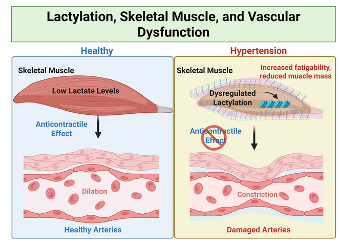
Emerging evidence suggests a critical interplay between skeletal muscle metabolism and vascular function in the context of hypertension. Elevated plasma lactate levels precede the onset of hypertension and are inversely associated with skeletal muscle mass, highlighting skeletal muscle atrophy and metabolic dysregulation as key contributors to cardiovascular dysfunction.
METHODS:
Male and female Wistar rats and spontaneously hypertensive rats were studied. Skeletal muscle performance was evaluated using in vivo plantarflexion torque measurements. Femoral arteries with surrounding skeletal muscle were isolated to assess contractility and relaxation. Plasma and muscle lactate levels were quantified using colorimetric assays. Structural remodeling and mitochondrial function were assessed via wheat germ agglutinin staining, succinate dehydrogenase activity, and high-resolution respirometry. Protein lactylation was evaluated by mass spectrometry-based lactylated proteomics. Human translational relevance was examined using publicly available skeletal muscle transcriptomic data.
RESULTS:
Spontaneously hypertensive rats exhibited skeletal muscle dysfunction marked by increased fatigability, reduced muscle mass, impaired mitochondrial activity, and elevated muscle lactate levels. Despite upregulation of oxidative markers, persistent lactate accumulation suggested a maladaptive metabolic shift. Proteomics revealed differential lactylation of key structural proteins (myosins, nebulin) and metabolic enzymes (Nampt, GAPDH [glyceraldehyde-3-phosphate dehydrogenase]). The anticontractile effect of skeletal muscle on femoral arteries was completely lost in spontaneously hypertensive rats, accompanied by impaired vascular relaxation and increased arterial lactylation. Human transcriptomic data supported parallel metabolic alterations in hypertension.
CONCLUSIONS:
Hypertension disrupts skeletal muscle metabolic homeostasis and muscle-vascular communication, driven in part by persistent lactate accumulation and altered protein lactylation. Targeting lactate-mediated signaling may offer new therapeutic avenues for hypertensive vascular dysfunction.
Hyatt et al.
Microcirculation— May 2026
OBJECTIVE:
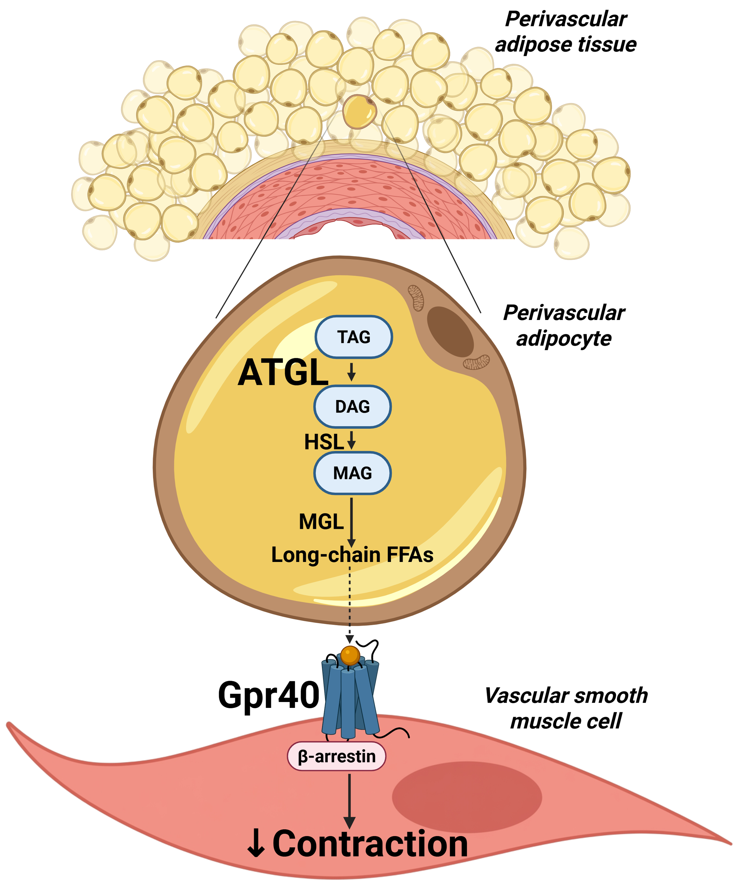
Perivascular adipose tissue (PVAT) surrounds most peripheral blood vessels and exerts an anti-contractile influence through paracrine mediators. Although numerous vasoactive factors have been identified, the mechanisms linking adipocyte metabolism to PVAT-dependent modulation of vascular tone remain poorly defined. Because adipocytes store energy as triglycerides hydrolyzed by adipose triglyceride lipase (ATGL) and hormone-sensitive lipase (HSL) to generate free fatty acids, we hypothesized that lipolysis-derived fatty acids may contribute to PVAT's anti-contractile actions through activation of long-chain fatty acid-sensing G protein-coupled receptors, Gpr40 and/or Gpr120.
METHODS:
Mesenteric resistance arteries (MRAs) from adult Wistar rats were studied using wire myography, with or without PVAT, and pharmacological agonists/antagonists and endothelial denudation were used to study signaling. To examine changes in hypertension, PVAT and MRAs from spontaneously hypertensive rats (SHRs) were analyzed by western blotting, and plasma from non-fasting or fasting SHR was assessed by untargeted lipidomics.
RESULTS:
In Wistar rats, inhibition of ATGL, but not HSL, abolished PVAT's anti-contractile effect, and blockade of Gpr40, but not Gpr120, similarly diminished this response, identifying ATGL and Gpr40 as important mediators. Activation of Gpr40 in PVAT- and endothelium-denuded MRAs further recapitulated the anti-contractile effect in a β-arrestin-dependent manner. In SHR, PVAT ATGL expression was significantly upregulated and MRA Gpr40 expression tended to increase. However, circulating Gpr40 ligand abundance was largely unchanged between strains, suggesting that impaired ligand availability is unlikely to underlie PVAT dysfunction in hypertension.
CONCLUSIONS:
These findings identify a previously unrecognized ATGL-Gpr40 signaling axis linking adipocyte triglyceride metabolism to PVAT-mediated regulation of vascular tone.
Nampoothiry et al.
Journal of Vascular Research — April 2026
INTRODUCTION:
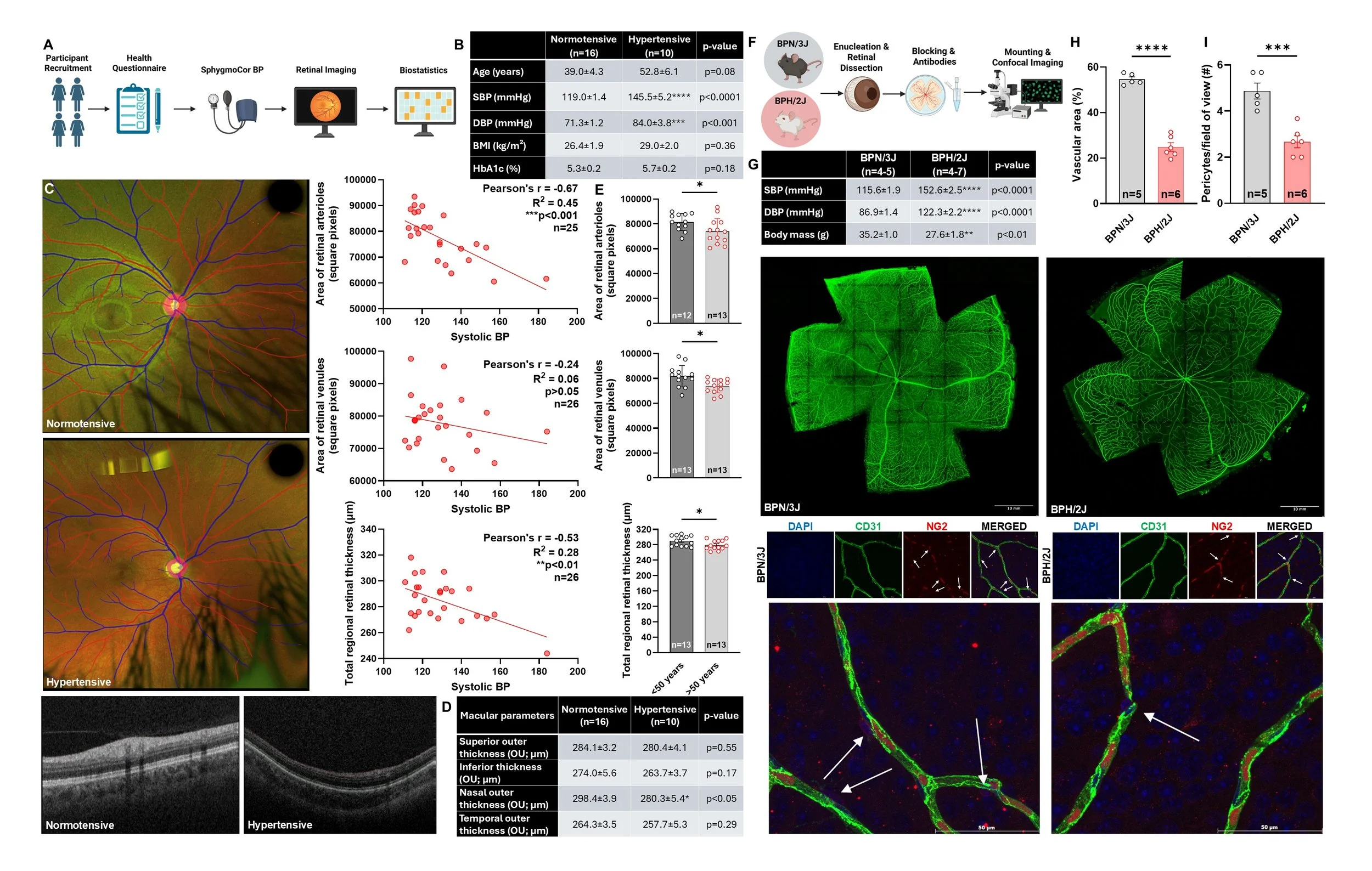
Hypertension is a major risk factor for cardiovascular disease, yet its effects on the retinal microvasculature in women, independent of pregnancy and diabetes, remain poorly understood. Methods: We integrated retinal imaging in female participants with structural assessments in hypertensive female mice to evaluate retinal microvascular remodeling.
METHODS:
We integrated retinal imaging in female participants with structural assessments in hypertensive female mice to evaluate retinal microvascular remodeling.
RESULTS:
In women, higher systolic blood pressure was associated with reduced retinal arteriolar area and retinal thinning, independent of BMI or glycemic status. Women over age 50 exhibited greater retinal microvascular loss, suggesting age-related susceptibility. After adjusting for age, retinal arteriolar area remained significantly associated with hypertension, whereas venular area and retinal thickness were primarily associated with age. No significant age × hypertension interactions were observed for any outcome. In parallel, hypertensive BPH/2J mice displayed retinal microvascular rarefaction and reduced pericyte coverage compared with normotensive BPN/3J controls.
CONCLUSION:
Across species, hypertension induced rarefaction, arteriolar loss, and pericyte depletion, indicating impaired microvascular integrity. These findings support the utility of retinal imaging as a noninvasive biomarker of hypertensive microvascular disease in women.
McCarthy et al.
American Journal of Physiology-Heart and Circulatory Physiology — May 2026
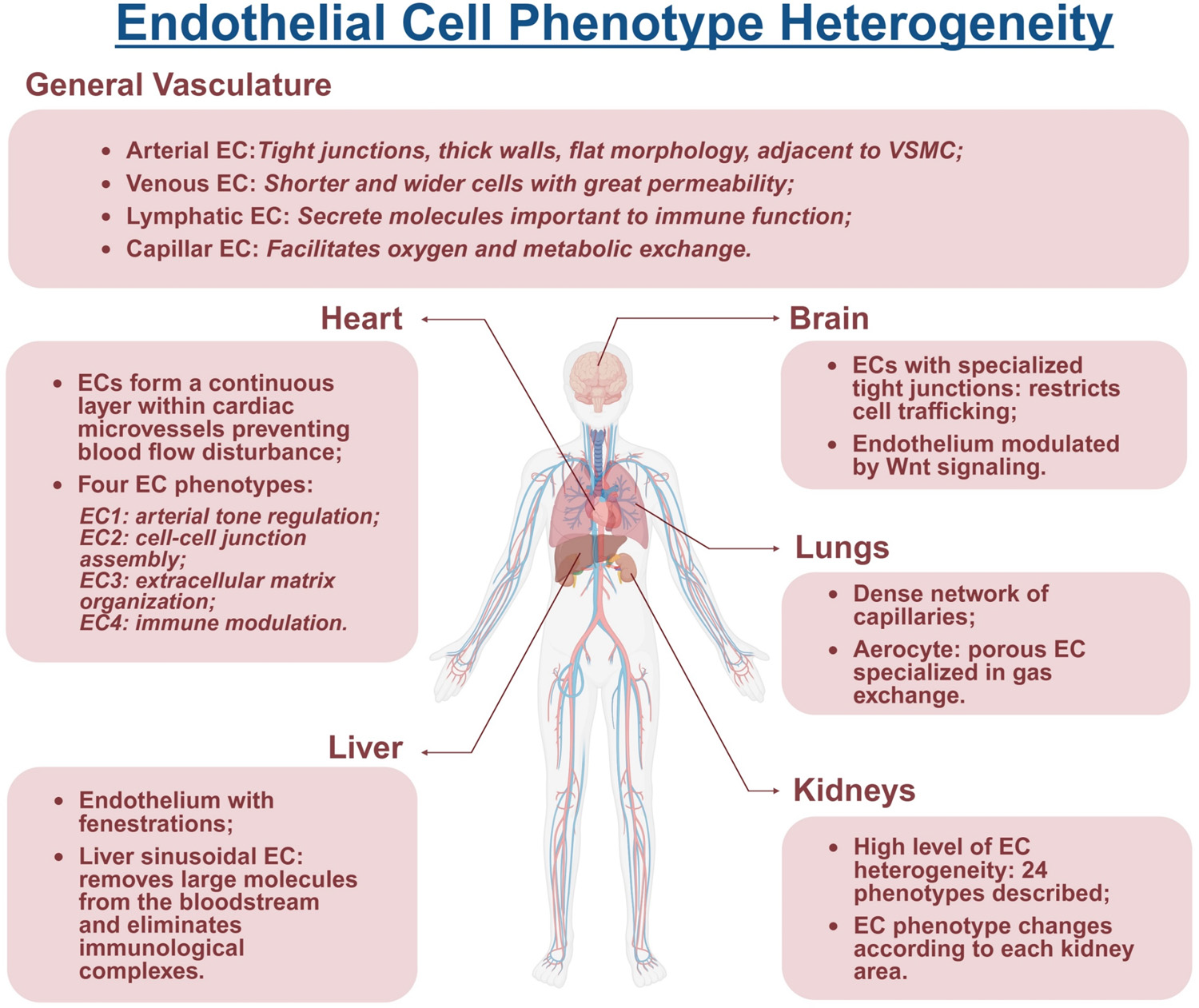
The endothelium plays a central role in maintaining vascular homeostasis by orchestrating vascular tone, inflammation, healing, permeability, and thrombosis. Assessing endothelial function in vascular tissue is essential for understanding the cellular and molecular mechanisms underlying cardiovascular physiology and pathology. Traditional approaches, such as wire and pressure myography, have been instrumental in defining endothelium-dependent responses and identifying key pharmacological targets. However, the complexity and heterogeneity of endothelial cells across vascular beds and their dynamic phenotypic changes in health and disease necessitate the incorporation of new investigative strategies. Emerging methodologies, including bulk and single-cell transcriptomics, proteomics, and advanced imaging, now provide unprecedented insights into endothelial cell diversity and function. A team of leading experts in the field, who collectively reached a consensus on the most widely used techniques to evaluate endothelial function, developed these guidelines. The document establishes best practices for assessing endothelial function, from endothelial cell cultures to isolated vascular tissues, integrating conventional functional assays with modern molecular approaches. By fostering methodological consistency and embracing innovation, our goal is to enhance rigor, reproducibility, understanding, and discovery in endothelial biology.
Listen to this article’s corresponding podcast featuring Dr. Wenceslau here!
Pernomian et al.
Circulation Research — September 2025

BACKGROUND:
Small artery remodeling and endothelial dysfunction are hallmarks of hypertension. Evidence supports a likely causal association between cardiovascular diseases and endothelial-to-mesenchymal transition, a cellular transdifferentiation process in which endothelial cells (ECs) partially lose their identity and acquire mesenchymal phenotypes. EC reprogramming represents an innovative strategy in regenerative medicine to prevent deleterious effects induced by cardiovascular diseases.
METHODS:
Using partial reprogramming of ECs, via overexpression of Oct-3/4-Sox-2-Klf-4 (OSK) transcription factors, we aimed to bring ECs back to a youthful phenotype in hypertension. Primary ECs were infected with lentiviral vectors (LVs) containing the specific EC promoter Cdh5 (cadherin-5) and the reporter EGFP (enhanced green fluorescent protein) with empty vector (LV control) or LV with Oct-3/4-Sox-2-Klf-4. Confocal microscopy and Western blotting analysis were used to confirm OSK overexpression. Cellular migration, senescence, and apoptosis were evaluated. Human aortic ECs from normotensive patients and patients with hypertension were analyzed after OSK treatments for eNOS (endothelial nitric oxide synthase), NO, and genetic profile. Male and female normotensive (blood pressure normal mouse strain) and hypertensive (blood pressure high mouse strain) mice were treated with LV control or LV with Oct-3/4-Sox-2-Klf-4 and evaluated 10 days post-infection. The blood pressure, cardiac function, vascular reactivity of small arteries, and endothelial-to-mesenchymal transition inhibition were analyzed.
RESULTS:
OSK overexpression induced partial EC reprogramming in vitro, and these cells had lower migratory capability. OSK treatment of blood pressure high mouse strain mice reduced blood pressure and resistance arteries hypercontractility, via the attenuation of endothelial-to-mesenchymal transition and elastin breaks. EGFP was detected in vivo in the prefrontal cortex. OSK-treated hypertensive human aortic ECs showed high eNOS activation and NO production, with low reactive oxygen species formation. Single-cell RNA analysis showed that OSK alleviated EC senescence and endothelial-to-mesenchymal transition, restoring their phenotypes in human aortic ECs from patients with hypertension.
CONCLUSIONS:
Overall, these data indicate that OSK treatment and EC reprogramming can decrease blood pressure and reverse hypertension–induced vascular damage.
2022
McCarthy CG, Waigi EW, Yeoh BS, Mell B, Vijay-Kumar M, Wenceslau CF, Joe B. Low-dose 1,3-butanediol reverses age-associated vascular dysfunction independent of ketone body β-hydroxybutyrate. Am J Physiol Heart Circ Physiol. 2022 Mar 1;322(3):H466-H473. doi: 10.1152/ajpheart.00486.2021. Epub 2022 Feb 11. PubMed PMID: 35148235; PubMed Central PMCID: PMC8897007.
Costa, Tiago J.a,b; Linder, Braxton A.c; Hester, Setha,b; Fontes, Milenea,b; Pernomian, Laenaa,b; Wenceslau, Camilla F.a,b; Robinson, Austin T.c; McCarthy, Cameron G.a,b. The janus face of ketone bodies in hypertension. Journal of Hypertension 40(11):p 2111-2119, November 2022. | DOI: 10.1097/HJH.0000000000003243
Santos CVD, Kerkhoff J, Tomazelli CA, Wenceslau CF, Sinhorin AP, de Jesus Rodrigues D, Carneiro FS, Bomfim GF. Vasoconstrictor and hemodynamic effects of a methanolic extract from Rhinella marina toad poison. Toxicon. 2022 Oct 30;218:57-65. doi: 10.1016/j.toxicon.2022.08.018. Epub 2022 Sep 14. PMID: 36113683; PMCID: PMC9832923.
McCarthy CG, Waigi EW, Singh G, Castaneda TR, Mell B, Chakraborty S, Wenceslau CF, Joe B. Physiologic, Metabolic, and Toxicologic Profile of 1,3-Butanediol. J Pharmacol Exp Ther. 2021 Nov;379(3):245-252. doi: 10.1124/jpet.121.000796. Epub 2021 Sep 14. PubMed PMID: 34521698; PubMed Central PMCID: PMC9164310.
Roy S, Edwards JM, Tomcho JC, Schreckenberger Z, Bearss NR, Zhang Y, Morgan EE, Cheng X, Spegele AC, Vijay-Kumar M, McCarthy CG, Koch LG, Joe B, Wenceslau CF. Intrinsic Exercise Capacity and Mitochondrial DNA Lead to Opposing Vascular-Associated Risks. Function (Oxf). 2021;2(1):zqaa029. doi: 10.1093/function/zqaa029. Epub 2020 Nov 3. PMID: 33363281; PMCID: PMC7749784.
McCarthy CG, Chakraborty S, Singh G, Yeoh BS, Schreckenberger ZJ, Singh A, Mell B, Bearss NR, Yang T, Cheng X, Vijay-Kumar M, Wenceslau CF, Joe B. Ketone body β-hydroxybutyrate is an autophagy-dependent vasodilator. JCI Insight. 2021 Oct 22;6(20). doi: 10.1172/jci.insight.149037. PubMed PMID: 34499623; PubMed Central PMCID: PMC8564907.
McCarthy CG, Saha P, Golonka RM, Wenceslau CF, Joe B, Vijay-Kumar M. Innate Immune Cells and Hypertension: Neutrophils and Neutrophil Extracellular Traps (NETs). Compr Physiol. 2021 Feb 12;11(1):1575-1589. doi: 10.1002/cphy.c200020. Review. PubMed PMID: 33577121; PubMed Central PMCID: PMC9721119.
Schreckenberger ZJ, Wenceslau CF, Joe B, McCarthy CG. Mitophagy in Hypertension-Associated Premature Vascular Aging. Am J Hypertens. 2020 Sep 10;33(9):804-812. doi: 10.1093/ajh/hpaa058. Review. PubMed PMID: 32533696; PubMed Central PMCID: PMC7481986.
Cheon S, Tomcho JC, Edwards JM, Bearss NR, Waigi E, Joe B, McCarthy CG, Wenceslau CF. Opioids Cause Sex-Specific Vascular Changes via Cofilin-Extracellular Signal-Regulated Kinase Signaling: Female Mice Present Higher Risk of Developing Morphine-Induced Vascular Dysfunction than Male Mice. J Vasc Res. 2021;58(6):392-402. doi: 10.1159/000517555. Epub 2021 Sep 14. PMID: 34521095; PMCID: PMC8612963.
Wenceslau CF, McCarthy CG, Earley S, England SK, Filosa JA, Goulopoulou S, Gutterman DD, Isakson BE, Kanagy NL, Martinez-Lemus LA, Sonkusare SK, Thakore P, Trask AJ, Watts SW & Webb RC (2021). Guidelines for the measurement of vascular function and structure in isolated arteries and veins. American Journal of Physiology-Heart and Circulatory Physiology; DOI: 10.1152/ajpheart.01021.2020.
Silva CBP, Elias-Oliveira J, McCarthy CG, Wenceslau CF, Carlos D, Tostes RC. Ethanol: striking the cardiovascular system by harming the gut microbiota. Am J Physiol Heart Circ Physiol. 2021 Aug 1;321(2):H275-H291. doi: 10.1152/ajpheart.00225.2021. Epub 2021 Jun 18. PMID: 34142885; PMCID: PMC8410123.
Edwards, J. M., Roy, S., Galla, S. L., Tomcho, J. C., Bearss, N. R., Waigi, E. W., Mell, B., Cheng, X., Saha, P., Vijay-Kumar, M., McCarthy, C. G., Joe, B., & Wenceslau, C. F. (2021). FPR-1 (Formyl peptide receptor-1) activation promotes spontaneous, premature hypertension in Dahl Salt-Sensitive rats. Hypertension, 77(4), 1191–1202. https://doi.org/10.1161/hypertensionaha.120.16237
2021
2020
2023
Waigi EW, Webb RC, Moss MA, Uline MJ, McCarthy CG, Wenceslau CF. Soluble and insoluble protein aggregates, endoplasmic reticulum stress, and vascular dysfunction in Alzheimer's disease and cardiovascular diseases. Geroscience. 2023 Jun;45(3):1411-1438. doi: 10.1007/s11357-023-00748-y. Epub 2023 Feb 24. PMID: 36823398; PMCID: PMC10400528.
Pernomian L, Tan W, McCarthy CG, Wenceslau CF. Reprogramming endothelial and vascular smooth muscle cells to prevent and treat hypertension. Medical Hypotheses [Internet]. 2023 Sep 7;179:111162. Available from: https://doi.org/10.1016/j.mehy.2023.111162